In pursuit of memory
To understand why memory declines with age, scientists must first figure out how the brain builds memories
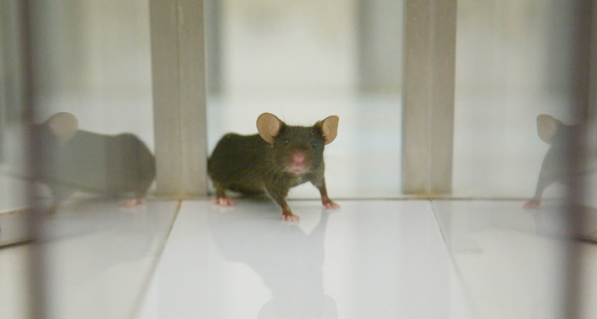
By studying mice like this one in the laboratory, researchers can peer into the mind and explore how memory works. Findings from experiments on lab mice might one day be used in therapies for people to slow or reverse some types of memory loss.
Alcino Silva
Some of the greatest discoveries in science have been made by accident, or chance. An important part of what scientists know about memory is among these.
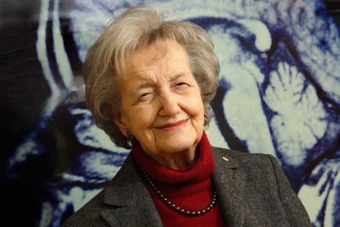
In the early 1950s, a young Canadian scientist named Brenda Milner earned a doctorate (Ph.D.) in neuroscience, or the study of the brain. Soon, she began working with a well-known surgeon, Wilder Penfield of the Montreal Neurological Institute in Canada. The doctor had developed a special technique to treat people with epilepsy. This disorder causes the brain to fire off electrical signals abnormally. These abnormal signals can trigger seizures, which often cause the body to shake, or convulse, violently. Seizures can be very severe, sometimes leading to further brain damage.
Penfield treated his patients by removing the brain region that was firing improperly. While this reduced seizures, two patients showed a deeply worrisome side effect: Shortly after surgery, they couldn’t remember their recent past. They didn’t even recognize their doctors, including Milner.
In 1953, Penfield presented these findings at a scientific meeting in Chicago. Soon after, he received a call from a neurosurgeon in Hartford, Conn. That doctor, too, had performed a brain operation on an epilepsy patient. And after the surgery, that patient — known only by his initials, H.M. (to protect his identity) — also could no longer recall memories of recent events. The surgeon invited Milner, a psychologist trained to study behavior, to come and study H.M.
Soon, she began taking a night train between Montreal and Connecticut regularly to study H.M.’s ability to learn and to build memories. “That was the beginning of the whole story,” Milner recalls.
Now 95, she still works at the Montreal Neurological Institute. Off and on for more than 50 years, Milner continued to study Henry Molaison — H.M.’s name was made public after his death in 2008. Her data confirmed that although removing the misfiring part of this man’s brain had controlled his seizures, he was never again able to form certain types of memories.
On tests of his IQ, or intelligence quotient, Molaison scored high. In fact, his IQ rating improved after the surgery. He also performed well on reasoning tests. And his active, or “short-term,” memory was fine. For instance, if someone gave him a telephone number, he could repeat it right back. But day-to-day, he never recognized Milner or the other researchers who worked closely with him.
This condition, called amnesia (am NEE she ah), is an inability to recall the past. It interferes with long-term recall. “This memory that is lost in these patients is so devastating because it’s what we call our autobiographical memory,” explains Milner. “It’s our memory of our life as we live it.”
Molaison was an important patient. What Milner and other scientists learned from studying him continues to shape neuroscience.
Scientists had once thought the whole brain controlled memory. But through the study of Molaison’s mind, they learned that all memories aren’t created the same way and in the same place. They now know that several different brain regions guide the building of memories.
“For the last 60 years, people have been trying to figure out what those brain areas do,” notes Mark Mayford, a neuroscientist. He works at the Scripps Research Institute in La Jolla, Calif.
Little by little, he and others have been piecing together an understanding of how memory works. But they are not even close to having a complete picture. “We don’t know much [about memory] with great certainty,” says Mayford. “We know a lot with sort of a little bit of certainty.”
Memories as mail
Molaison’s surgery had removed most of an area of the brain called the hippocampus. This region consists of two long ridges that sit deep within the brain, about halfway between the eyes and the back of the head. Because Molaison went on to have trouble making new memories, scientists now know that this area is critical for forming them.
Fred Gage, a neuroscientist at the Salk Institute for Biological Studies, also in La Jolla, offers one example. Imagine you are in class, listening to your teacher discuss a difficult aspect of math. You’re hearing something new — and hopefully learning. In addition, “You’re seeing, smelling the person next to you a little bit, you’re hungry, and you’re slightly distracted by the fact that you’re late with your mid-term paper,” says Gage. All of this sensory information — smells, sounds, worries, hunger pangs and new learning — piles onto a set of brain cells, called neurons, in the temporal lobe. It contains the hippocampus.
All of the incoming scattered bits of sensory information pass through a circuit — like a pipeline — into the hippocampus. There, a special processor with a strange name, the dentate gyrus, sorts through all the information. It determines what’s new and what isn’t; which events are related, and which aren’t.
Then, all of those individual, messy pieces of data are stored together as one package in yet another part of the hippocampus. Think of this step as tying a bunch of loose pieces of mail into one bundle. This new package of information gets shipped out of the hippocampus and delivered back to the outermost layer of the brain. Called the cortex, this layer is like a watermelon rind. That newly formed package is a memory. But “how it’s distributed, and where in the cortex is not so clear,” notes Gage.
When you later recall what you learned in math class that day, you might also recall the scent of the perfume worn by the girl who was sitting next to you. That’s because such bits of sensory information were bundled into the same memory. This is called an association.
Associations help explain what happens in the brains of people who have endured a traumatic event, such as those with post-traumatic stress disorder. Troops who have survived explosions or other war traumas sometimes develop this PTSD. For instance, a soldier might have been driving a vehicle and listening to horns honking right when a bomb went off. Years later, a honking horn can trigger severe anxiety or recollections of the explosion. The sound alone can bring back vivid memories of the earlier near-death experience.
Two types of memories
There are two key types of memories: episodic (memories of events, or episodes) and procedural. When you listened to the math lesson and at the same time worried about a big writing assignment that was due soon, you formed an episodic memory. When you learned how to tie your shoelaces, ride a bike or pop open a soda can, you formed a procedural memory.

Molaison could make procedural memories, however. Such memories are formed in a part of the brain not touched during his surgery. Milner remembers the moment she discovered her patient could form these types of memories as the most exciting in her career.
She had given Molaison a pencil and a drawing of a star with a double border. Then Milner had asked him to draw a line around the star, staying within the border. But Molaison was not allowed to look directly at the star on the paper — he had to look at its reflection in a mirror. Called mirror drawing, scientists use this task to test peoples’ motor-learning skills (here, motor refers to movement). The reflection can mislead the eye, making it difficult to follow the actual pattern.
Milner allowed Molaison to perform the task 10 times a day for three days. Each time, he got better. Finally, on the very last trial, he traced the star perfectly between the lines. Afterward, the scientist recalls, “He stood up and he said, ‘Well, that’s funny — I thought that was going to be difficult, but it looks as though I’ve done it rather well.’”
Molaison was totally unaware that he had performed the task nearly 30 times before. So he “was justifiably very proud of himself,” Milner explains. His ability to improve until he had perfected the task proved that he could build procedural memories — even though he couldn’t remember having done so.
Making —and missing —connections
Understanding exactly how various types of memories form — and how we recall them — is proving tricky. But in the last few years, brain scientists have made exciting advances. “We now have for the first time the building blocks of memory, which we couldn’t say even five years ago,” notes Alcino Silva. He is a neuroscientist at the University of California, Los Angeles.
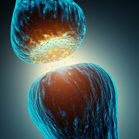
Silva studies how synapses (SIN ap seez) function — or act — in the brain. These tiny structures are like bridges that link nerve cells. Nerve cells send chemical and electrical signals to each other across synapses. When learning is underway, or memories are forming, these bridges get stronger and stronger. Their strengthening is needed for memory creation and recall, explains Silva. “We now have overwhelming evidence that during learning, synapses change and that these changes are absolutely essential to our brain’s ability to learn.”
When a learning connection, or memory, occurs in the brain, a message will move to its intended recipient. The next time your brain is presented with a similar problem, the message will be transmitted more efficiently. That’s because having dealt with that type of problem before, it has learned what to do. Brain scientists call the network built through this process the brain’s scaffolding. It provides a structural support for memories, just as scaffolds — or framing — provide the skeleton for a building.
The brain’s bridges frequently don’t form properly, though. When one nerve cell sends an electrical impulse to another, that signal often is not received. Think of it like a relay race. One runner has to go a certain distance, then pass a baton, or stick, to the next runner. But imagine that during the hand-off, the baton drops. That’s essentially what happens when a nerve cell fires off a signal that its intended target fails to receive.
“It’s really amazing, but the brain is not made out of connections that work at high efficiency,” says Silva. In fact, he notes, “Most connections in the brain between brain cells actually work at very low efficiency. One cell sends a signal and the signal is lost. The other cell does not respond at all, most of the time.”
The more you learn and the more memories you build, however, the better you are able to learn and increase nerve cell communication. The runner now seldom drops the baton. Instead, she races forward and successfully hands it off.
Red light, green light
The failure of nerve cells to relay signals properly underlies some learning disorders. Silva studies such learning problems in mice and other animals. By studying those animals, scientists can better understand the human brain. Silva’s team is finding that learning problems occur when messages get mixed up during transmission.
Sometimes a cell in the message relay is incorrectly told to stop. That’s like hitting the brakes in your car when you should be stepping on the accelerator, explains Silva. For example, he says, take Los Angeles’ traffic lights. If they were always red, no driver could advance through an intersection. And if they were always green, everybody would fight to go at once and create a traffic jam. “The brain is the same. It’s a balance between stop and go,” Silva says. His mice can’t learn new tasks very well because instead of giving a green light to the appropriate brain cell, “the brain is telling it ‘Stop! Stop!’” This keeps the connections that are needed for learning from being built and strengthened.
But there are encouraging signs for treatment of such learning problems in mice — and people. Silva’s team has found that injecting mice with certain drugs removes the brake on brain-signal transmissions. That brake is actually a messenger chemical called GABA. Some drugs can slow or stop the brain’s release of GABA. So instead of receiving a red light, treated nerve cells now receive a green one. Mice receiving this GABA blocker can suddenly learn normally.
Now, Silva and others are performing trials to test whether this drug will also work for memory problems in people.
Altered memories
Another discovery that has fascinated scientists is that past memories often change, becoming different each time we recall them.
“Our memory is constructive,” says Denise Park. “We often construct things to make sense of a memory that is only partially recalled. One result: Some of what we think we remember is not entirely true,” she explains. Park is a neuroscientist at the University of Texas in Dallas.
Over time, as memories become fuzzier, we automatically fill in the gaps with details. Although those details will seem reasonable, they may not be entirely accurate. Often, when we recall an old memory, we alter it slightly, then store it back away. It’s a bit like opening a file on a computer, making a few changes to it, then saving it.
As people age, these alterations happen more and more. Older adults have trouble remembering details and the time, place or context in which things occurred. For example, maybe you remember who you had lunch with, but not when or where. This is especially likely to occur as certain areas in the brain, including the hippocampus, shrink with age.
Park studies the aging brain. She is particularly interested in changes that occur in people during their 30s, 40s and 50s. She believes these midlife brain changes hold important clues to why some peoples’ brains age well while memory declines faster and more severely in others.
To study this, she peers inside the brain using something called functional magnetic resonance imaging, or fMRI. This technology allows scientists to watch what is happening in the brain in real-time — as people are learning, thinking and remembering. Researchers can watch tens of thousands of nerve cells light up every couple of seconds as an individual reads, listens or views something. Those images point to cells that were active and working together.
Such advances were almost unimaginable when Milner began her career. “In those faraway days, we didn’t have a good way of looking into the brain,” she says. “You just got a picture — an X-ray of the skull and the shape of [big structures] of the brain. But you did not get any idea what was happening.”
Today, she says, imaging the living brain is transforming not only how scientists design experiments but also what they can learn about the mind. “We can see the normal living brain in action,” she explains. In 1953, the idea of doing that would have been “just magical thinking.”
Scientists hope it might be possible one day to fix age-related memory problems and memory losses caused by disease — or to prevent them altogether. But first, they have to get a fuller picture of the processes involved in memory and learning, says Mayford. “Until we understand how the brain is functioning, it becomes hard to understand how these things go wrong.”
Power Words
Alzheimer’s disease A form of brain decline that gets worse over time and that affects memory, thinking and behavior.
amnesia Impaired long-term memory caused by brain damage, disease, shock or trauma.
cortex The outermost layer of neural tissue of the brain.
dentate gyrus Part of the hippocampal region, thought to contribute to the formation of new episodic memories.
epilepsy A neurological disorder characterized by seizures.
episodic memory The memory of past experiences, including times, places, people, emotions, knowledge and other information about the world.
functional magnetic resonance imaging (fMRI) An imaging technique used to visualize internal structures of the body. Whereas regular MRI allows imaging of the brain as still shots — like an X-ray — fMRI allows scientists to study neural activity in real-time.
hippocampus Elongated ridges found on each side of the brain. They are thought to be the center of emotion, memory and the involuntary nervous system.
GABA An abbreviation for gamma-aminobutyric acid. This chemical messenger acts as a brake, or inhibitor, on the firing of brain cells.
IQ, or intelligence quotient A number representing a person’s reasoning ability. It’s determined by dividing a person’s score on a special test by his or her age, then multiplying by 100.
long-term memory The brain’s system for storing, maintaining and recalling information from the past.
motor skills Ability to make controlled movements with the hands or limbs.
neuron, or nerve cell Any of the impulse-conducting cells that make up the brain, spinal column and nervous system.
post-traumatic stress disorder (PTSD) A severe condition that may develop after a person is exposed to a traumatic event. Recalling the event can bring on anxiety and other problems in the recaller.
short-term memory (also known as primary memory) The small amount of memory held actively in the mind for a short period of time, such as the series of digits in a telephone number.
synapse The junction, or bridge, between neurons that transmits chemical and electrical signals.
temporal lobe A regionof the brain where long-term memories are formed.
