Memory lessons from a forgetful brain
Scientists have just begun probing the preserved tissue from a man who, following brain surgery, suffered memory problems for the rest of his life

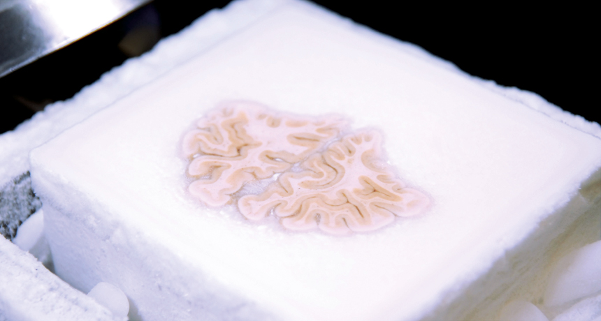
After being frozen solid, Henry Molaison’s brain (shown here) would be sliced into tissue-paper-thin wedges and then photographed.
Annese et al.
In 1953, a brain surgeon accidentally took away Henry Molaison’s ability to make new memories. The patient was only 27 years old. But Molaison’s loss became a major gain for science. For the rest of his life, this man — long referred to only as H.M. — donated much of his time to scientists. He wanted them to learn from his tragedy. When Molaison died in 2008, he donated his brain to science. And a new analysis of it is offering memory experts additional lessons.
This study finds that the basis for Molaison’s persistent memory loss appears more complicated than scientists initially had thought.
As a young man, Molaison suffered from severe and frequent seizures. To ease them, surgeon William Beecher Scoville removed parts of the man’s brain. The procedure worked — but had a major, unintended side effect: Molaison became unable to form new memories. Scientists realized they could link this problem to his drastically altered brain. Before long, scientists began publishing findings they were gleaning from their study of H.M.
Indeed, “The studies on H.M. began the present era of research on how the brain supports memory,” notes Howard Eichenbaum. A neuroscientist at Boston University, he studies how the brain and other parts of the nervous system work.
Recently, Jacopo Annese of the Brain Observatory at the University of California, San Diego and his colleagues decided to study Molaison’s donated brain. Their technique provides more detail than anything that can be used on a living patient. It required freezing the brain and then slicing it into 2,401 paper-thin wisps.
Scientists scanned a photograph of each slice into a computer. A computer program then reassembled the images — virtually — to create a 3-D model of that brain. This allowed Annese’s team to view every part and from any angle, inside and out.
Then these researchers compared this model to drawings that Molaison’s surgeon had made shortly after operating on the man’s brain. The hippocampus is one of the primary memory-forming structures in the brain. Scoville had indicated he had removed the entire hippocampus. The new 3-D computer model shows that the surgeon actually left behind a surprising amount of the hippocampus.
This wasn’t a total surprise. Brain scans from when Molaison was alive had shown that some of the hippocampus remained. But the amount seen in those brain scans was much less than the 3-D computer reconstruction now finds. Moreover, what hippocampus tissue Molaison retained still looked healthy when he died. Annese’s team reported its findings Jan. 28 in the research journal Nature Communications.
These data indicate that Molaison’s severe memory problems are not solely due to a missing hippocampus, notes Eichenbaum, who wasn’t involved in the study. The loss of brain structures in addition to the hippocampus likely also played a role, he says.
For instance, the surgery removed a brain region called the entorhinal (EN toh RYE nul) cortex. It relays signals between the hippocampus and the rest of the brain. The loss of this entorhinal cortex probably had a big role in Molaison’s memory problems, Eichenbaum concludes.
And knowing that may prove important, he says. Why? Comparing Molaison’s memory problems with those of people who lack just the hippocampus might help scientists tease apart the jobs of the various brain structures involved in memory, he says.
Power Words
computer model A program that runs on a computer that creates a model, or simulation, of a real-world feature, phenomenon or event.
entorhinal cortex A portion of the brain that serves as a relay station to transmit signals between the hippocampus (memory center) and the rest of the brain.
hippocampus A seahorse-shaped region of the brain. It is thought to be the center of emotion, memory and the involuntary nervous system.
nervous system The network of nerve cells and fibers that transmits signals between parts of the body.
neuroscience Science that deals with the structure or function of the brain and other parts of the nervous system. Researchers in this field are known as neuroscientists.
computer program A set of instructions that a computer uses to perform some analysis or computation.
seizure A sudden surge of electrical activity within the brain. Seizures are often a symptom of epilepsy and may cause dramatic spasming of muscles.
virtual Being almost like something. Something that is virtually real would be almost true or real — but not quite. The term often is used to refer to something that has been modeled by or accomplished by a computer using numbers, not by using real-world parts. So a virtual motor would be one that could be seen on a computer screen and tested by computer programming (but it wouldn’t be a three-dimensional device made from metal).


