Teeny tiny hairs on brain cells could have big jobs
These small spikes could limit weight gain or help cells communicate

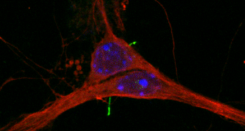
Mouse nerve cells (red) each sport a hairlike stub. It’s called a primary cilium (green). These tiny antennae may have many roles in the brain.
Ruchi Bansal and N. Berbari
Most cells in the body — including those in the brain — have a single tiny antenna. These short, narrow spikes are known as primary cilia (SILL-ee-uh). Each one is made of fat and protein. And these cilia will have different jobs, depending on where their host cells live. In the nose, for example, these cilia detect odors. In the eye, they help with vision. But their role in the brain has remained largely a mystery. Until now.
There are no odors to smell or light to see in the brain. Still, those tiny stubs appear to have big jobs, a new study reports. For instance, they may help control appetite — and possibly obesity. These cilia seem to contribute to brain development and memory. They might even help nerve cells chat.
“Perhaps every neuron in the brain possesses cilia,” says Kirk Mykytyn. Yet, he adds, most people who study the brain don’t even know they’re there. Mykytyn is a cell biologist. He works at Ohio State University College of Medicine in Columbus.
Christian Vaisse is a molecular geneticist. That’s someone who studies the role of genes — bits of DNA that give instructions to a cell. He is part of a team at the University of California, San Francisco who studied a protein called MC4R in search of clues about what cilia might do in the brain.
His group had known that tiny changes in the way MC4R does its job could lead to obesity in people. In mice, MC4R is made in the middle of the cell. Later, it moves to take up residence on cilia of the brain cells that help control mousey appetites. Vaisse and his colleagues already knew that MC4R didn’t always look the same. Some of its molecules looked unusual. The DNA in some cells must have developed some natural tweak — or mutation — that altered how the body made this protein.
Such mutations might also have changed how the protein worked.
For instance, one altered form of MC4R is connected to obesity. And in the mouse nerve cells making it, this form of the protein no longer shows up in the cilia where it belongs. When the scientists looked in the brain of a mouse with this mutation, they found, again, that MC4R was not on the nerve cell cilia where it should go to work.
The researchers then homed in on a different molecule, one that normally partners with MC4R. This second protein is called ADCY3. When they messed with it, it no longer cooperated with MC4R. Mice making these weird, lonely proteins also gained weight.
This may mean that MC4R needs to reach the cilia and dance with ADCY3 to work. Vaisse and his colleagues published this assessment on January 8 in the journal Nature Genetics.
From food to feelings
Researchers already knew that some unusual version of the MC4R protein was linked with obesity. Now, they have linked obesity to problems with the ADCY3 gene. Two studies on this were also published on January 8 in Nature Genetics. Both of these proteins work only once they climb aboard cilia. That new knowledge provides more support for the idea that cilia are involved in obesity.
These new studies aren’t the only clues linking cilia and obesity. A mutation that alters cilia also causes a very rare genetic disease in people. Obesity is one of its symptoms. The new findings hint that abnormal (mutant) cilia may play a role in obesity. And this may be true even in people without the genetic disease.
It’s also possible that other genes linked to obesity might need these cilia to do their work, Vaisse says.
Although data show the MC4R protein must reach cilia to control appetite, Mykytyn points out that no one knows why. It’s possible that the hairlike extensions have the right mix of helper proteins to let MC4R control appetite. Cilia might also change the way the protein works, perhaps making it more efficient.
Clearly, questions remain. Still, the new study “opens up the window a little more” on what cilia actually do in the brain, says Nick Berbari. He says it shows some of the things those cilia do — and what can happen when they don’t get their jobs done. Berbari is a cell biologist in Indianapolis at Indiana University-Purdue University.
Sending brain cell mail
Dopamine (DOPE-uh-meen) is a vital chemical in the brain that serves as a signal to relay messages between cells. Mykytyn and his colleagues have turned up a protein in cilia that detects dopamine. This sensor needs to be on cilia to do its work. Here, cilia might serve as a cell’s antenna, waiting to catch dopamine messages.
The stubby antennae may even be able to send cell-mail themselves. That was first reported in a 2014 study. They were studying nerve-cell cilia in worms known as C. elegans. And those cilia could dispatch little chemical packets into the space between cells. Those chemical signals may have a role in the worms’ behavior. The scientists published their worm study in the journal Current Biology.
Cilia also may have roles in memory and learning, Berbari says. Mice lacking normal cilia in parts of their brain that were important for memory had trouble remembering a painful shock. These mice also didn’t recognize objects as well as did those with normal cilia. These findings suggest mice need healthy cilia for normal memories. Berbari and his colleagues published those findings in 2014 in the journal PLOS ONE.
Finding out just what cilia do in the brain is a tough job, Mykytyn says. But new tricks in microscopy and genetics may reveal more about how these “underappreciated appendages” work, Berbari says. Even in places as busy as the brain.


